Vestibular balance rehabilitation therapy (VRT) uses specialized exercises to reduce dizziness, improve gaze stability, and restore postural control in patients with vestibular system dysfunction. Clinical evidence consistently supports VRT as the first-line non-pharmacological treatment for peripheral vestibular disorders, and clinicians can achieve measurable outcomes across etiologies when they match protocols to patient presentation.
Clinical Rationale: Why Vestibular Rehabilitation Works
VRT produces functional improvement through three distinct neurophysiological mechanisms: adaptation, substitution, and habituation. Vestibular adaptation refers to long-term changes in the gain of the vestibulo-ocular reflex (VOR), driven by retinal slip error signals that prompt the cerebellum to recalibrate central processing. Substitution recruits alternative sensory systems—somatosensory input, cervical proprioception, and visual flow—to compensate for lost peripheral vestibular function. Habituation reduces symptomatic responses to provocative stimuli through repeated, controlled exposure that progressively desensitizes central pathways.
A foundational PMC review (PMC3259492) confirms that vestibular compensation is an active, use-dependent process that stalls without targeted exercise. This is clinically significant: patients who rest to avoid symptoms suppress the very sensory conflict signals the CNS requires to recalibrate. Pharmacological vestibular suppressants, while appropriate for acute symptom management, should be tapered before initiating VRT to avoid blunting adaptive signals. The Vestibular Disorders Association (VEDA) identifies VRT as effective for the majority of vestibular disorders when initiated at the appropriate phase of recovery.
Patient Stratification: Who Is a Candidate for VRT
Appropriate candidate selection determines whether VRT produces meaningful outcomes or stalls due to mismatched protocol design. Strong candidates include patients with unilateral vestibular hypofunction (UVH) secondary to vestibular neuritis or labyrinthitis, post-acoustic neuroma resection, bilateral vestibular hypofunction, and chronic BPPV residual symptoms following successful canalith repositioning procedures for BPPV. Patients with compensated Ménière’s disease between active hydrops episodes also benefit from structured balance retraining.
Exclusion or deferral criteria include active BPPV (which requires repositioning first, not habituation), acute Ménière’s attacks, and uncontrolled central pathology such as progressive cerebellar degeneration or acute brainstem lesions. Clinicians should complete vestibular assessment tools used in clinical practice before initiating any protocol to establish baseline compensation status and etiology.
Peripheral vs. Central Vestibular Disorders: Differential Considerations
Peripheral vestibular disorders—including labyrinthitis, vestibular neuritis, and post-resection acoustic neuroma—typically present with unidirectional nystagmus, positive head impulse test, and symptoms that fatigue with sustained provocation. These patients respond well to adaptation-based VOR training and sensory reweighting. Ménière’s disease introduces a fluctuating hypofunction model that complicates static protocol design; clinicians should prioritize fall prevention and postural stability during quiescent phases rather than aggressive VOR gain training.
Central vestibular disorders require careful screening before VRT initiation. Red flags include direction-changing nystagmus, skew deviation, dysmetria, ataxia disproportionate to reported dizziness, and acute onset headache. Patients presenting with these signs warrant immediate referral to neurology or neuro-otology before any exercise program begins. Refer to the differential diagnosis of balance disorders framework to guide triage decisions when etiology is unclear.
Core Protocol Domains: Gaze Stabilization, Habituation, and Balance Retraining
VRT protocols organize into three functional domains: gaze stabilization, habituation, and balance retraining. Each domain targets a distinct mechanism and requires specific dosing parameters to drive neuroplastic change. Cleveland Clinic describes VRT as a customized program requiring individualized progression, a position supported by the evidence base showing that generic exercise sets produce inferior outcomes compared to impairment-matched protocols. Clinicians should reassess patients every two to four weeks and adjust dosing based on symptom trajectory and objective performance data.
Gaze Stabilization Exercises: VOR x1 and x2 Protocols
Gaze stabilization exercises directly train VOR gain through controlled head-eye coordination tasks. The VOR x1 protocol instructs the patient to fix gaze on a stationary target while moving the head in the horizontal or vertical plane at a frequency of 1–2 Hz. Target distance should be 18–24 inches initially, with progression to 6 feet as gain improves. Sessions begin at 1–2 minutes per plane and progress to 5 minutes as tolerance allows.
The VOR x2 protocol introduces a moving target that travels in the opposite direction of head movement, doubling the retinal slip signal and accelerating adaptive gain changes. This protocol is reserved for patients who have plateaued on x1 or who present with documented low VOR gain on video head impulse testing (vHIT). Frequency targets of 1.5–2.5 Hz are appropriate for most patients, with progression benchmarked against symptom reduction and vHIT gain normalization toward 0.8 or above. Both protocols should be performed in two to three daily sessions of 5–20 minutes total, consistent with adaptation training literature.
Habituation vs. Adaptation: Choosing the Right Strategy
Habituation and adaptation are not interchangeable strategies. Clinicians should use habituation for patients who experience reproducible dizziness with specific movements or visual stimuli and who retain some peripheral vestibular function. The mechanism is repeated symptom provocation at a sub-maximal intensity, which progressively reduces the magnitude of the central response. Clinicians should use the Motion Sensitivity Quotient or a structured movement inventory to identify provocative positions and grade exposure systematically.
Adaptation protocols are the correct choice when the goal is VOR gain recalibration—specifically in patients with documented unilateral or bilateral hypofunction and insufficient spontaneous compensation. Patients with chronic, uncompensated UVH (greater than three months post-onset with persistent oscillopsia or gait instability) are the primary adaptation candidates. Combining both strategies in the same session is appropriate only after the patient demonstrates stable tolerance to habituation stimuli, as premature adaptation loading can exacerbate symptoms and reduce adherence.
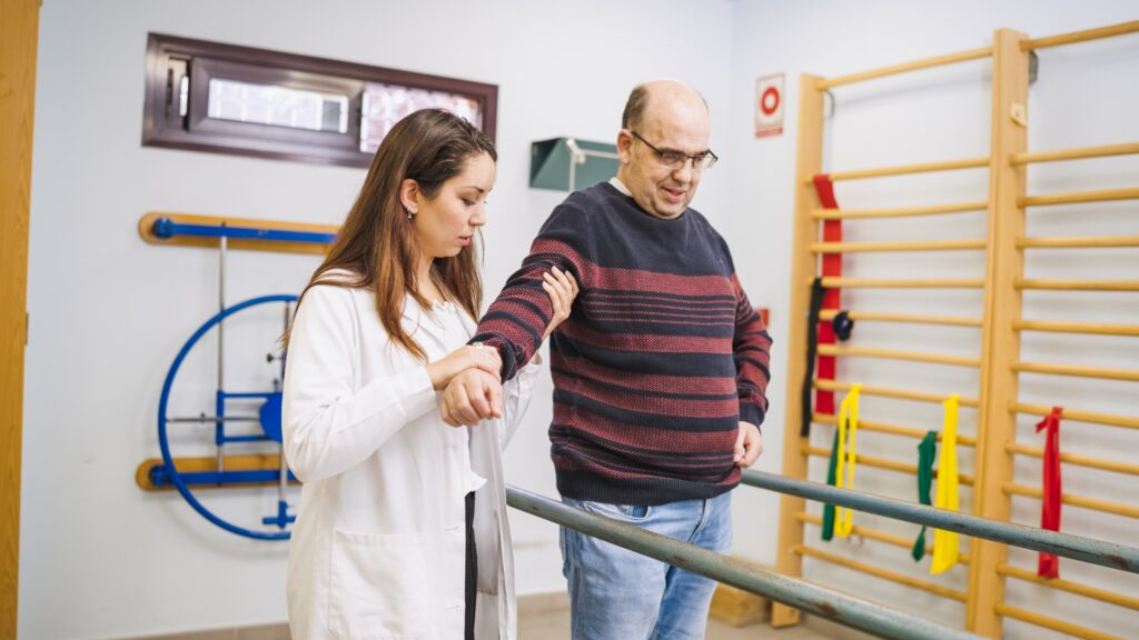
Balance Retraining: Sensory Reweighting and Postural Control Progressions
Balance retraining targets the CNS’s ability to appropriately weight and reweight sensory inputs—vestibular, visual, and somatosensory—under varying environmental conditions. NYU Langone describes sensory coordination training as a core component of vestibular rehabilitation for vertigo, particularly for patients with visual dependency or surface instability. The sensory organization test (SOT) from computerized dynamic posturography provides objective baseline data on which sensory conditions are most impaired and should directly inform exercise selection.
Progressions follow a foam-surface and visual conflict hierarchy: stable surface with eyes open → stable surface with eyes closed → foam surface with eyes open → foam surface with eyes closed → foam surface with visual conflict (e.g., optokinetic stimulus or busy visual environment). Clinicians introduce dual-task progressions, including cognitive load such as counting backward or word fluency tasks, after the patient achieves stable single-task performance on foam surfaces. They assess fall risk at each progression step and use gait belts or parallel bars for patients with DGI scores below 19/24 during training sessions.

Outcome Measurement Tools Every Vestibular Specialist Should Use
Clinicians need objective outcome measurement for evidence-based discharge planning and payer justification. Most VRT teams use the Dizziness Handicap Inventory (DHI) as the primary patient-reported outcome, with a minimum clinically important difference (MCID) of 18 points on the 100-point scale. Clinicians should complete detailed Dizziness Handicap Inventory scoring and interpretation at intake, four weeks, and discharge. The Dynamic Gait Index (DGI) assesses gait adaptability across eight conditions, and scores below 19/24 indicate elevated fall risk. The MiniBESTest (28-point scale) assesses broader postural control across anticipatory, reactive, sensory orientation, and dynamic gait domains; researchers have established an MCID of 4 points for vestibular populations. Clinicians often underuse the Pittsburgh Sleep Quality Index (PSQI) in VRT, but sleep disruption correlates with slower vestibular compensation, so teams should track it as a secondary outcome measure. Administer all four tools at baseline and reassess every four weeks to guide progression and establish safe discharge readiness confidently.
Treatment Timeline and Prognosis: Setting Evidence-Based Expectations
Vestibular rehabilitation typically produces measurable improvement in 6–8 weeks for unilateral peripheral disorders such as labyrinthitis or neuritis. Bilateral hypofunction and central vestibular conditions may require 12 or more weeks of structured therapy. Prognosis depends on etiology, chronicity, patient adherence, and baseline compensation status assessed at intake.
Additional prognostic variables include age, anxiety comorbidity, and medication burden. Patients taking benzodiazepines or antihistamines throughout the treatment course show slower compensation rates. Anxiety and somatosensory amplification, common in persistent postural-perceptual dizziness (PPPD), extend timelines and may require concurrent cognitive behavioral therapy. Clinicians should communicate disorder-specific timelines at the first visit to calibrate patient expectations and improve adherence.
Telehealth and Home Program Considerations for VRT
Home exercise programs (HEPs) are a structural component of VRT, not an adjunct. Frequency of exercise—ideally two to three sessions daily—drives adaptation more than session duration. Telehealth delivery of VRT has demonstrated comparable outcomes to in-person care for appropriate candidates, as supported by emerging literature on telehealth delivery of physical therapy home programs.
Safety screening for unsupervised home practice should include fall risk stratification (DGI, Timed Up and Go), home environment hazard review, and caregiver availability assessment for high-fall-risk patients. Adherence monitoring via exercise logs, app-based check-ins, or brief telehealth visits at weeks two and four significantly improves completion rates. Exercises requiring visual conflict or foam surfaces should not be assigned for unsupervised home practice until the patient has demonstrated safe performance in the clinic.
Clinical Pitfalls and When to Refer or Escalate
The most common clinical error in VRT is over-provoking symptoms during the initial phase, leading to patient avoidance and dropout. Symptom provocation should be graded: a 3–4/10 dizziness response is target intensity; responses above 6/10 indicate the exercise load exceeds current compensation capacity and must be reduced. A second frequent error is failing to progress the program, leaving patients on the same exercise set for weeks without objective reassessment.
Missing central pathology is the highest-stakes error. Any patient who fails to show measurable improvement after four weeks of correctly dosed VRT warrants re-evaluation for central etiology, including MRI referral if not already completed. Referral thresholds to neurotology include: persistent direction-changing nystagmus, progressive hearing loss concurrent with vestibular symptoms, suspected endolymphatic hydrops, or post-surgical complications. Neuro-ophthalmology referral is appropriate when ocular motor dysfunction—nystagmus, saccadic intrusions, or pursuit deficits—is disproportionate to the vestibular diagnosis.

FAQs
1. What exercises do you do for vestibular rehab and how are they dosed clinically?
VRT includes three primary exercise categories: gaze stabilization (VOR x1 and x2 protocols at 1–2.5 Hz, 5–20 minutes daily), habituation exercises targeting reproducible provocative movements, and balance retraining progressions from stable to foam surfaces with visual conflict. Dosing is individualized based on impairment profile, with two to three daily sessions recommended to drive neuroplastic adaptation.
2. Can vestibular rehabilitation therapy improve balance, and what outcome measures confirm improvement?
Yes. VRT consistently improves postural control through sensory reweighting and balance retraining. Improvement is confirmed using the MiniBESTest (MCID: 4 points), Dynamic Gait Index (fall risk threshold: 19/24), and the DHI (MCID: 18 points). Computerized dynamic posturography SOT scores provide objective sensory organization data before and after treatment.
3. How long does it take to treat vestibular balance disorders, and what variables affect prognosis?
Unilateral peripheral disorders typically resolve within 6–8 weeks of structured VRT. Bilateral hypofunction and central disorders require 12 or more weeks. Prognosis is affected by etiology, symptom chronicity, vestibular suppressant use, anxiety comorbidity, and patient adherence to the home exercise program.
4. How do clinicians differentiate between habituation and adaptation protocols for individual patients?
Habituation is appropriate for patients with reproducible, movement-triggered dizziness who retain some vestibular function. Adaptation is indicated for patients with documented VOR hypofunction and insufficient spontaneous compensation, particularly those with chronic UVH. The two strategies can be combined once habituation tolerance is established.
5. When is vestibular rehabilitation contraindicated or insufficient without medical co-management?
VRT should not be initiated during active BPPV (repositioning first), acute Ménière’s attacks, or in the presence of central red flags such as direction-changing nystagmus or progressive ataxia. Patients with PPPD, significant anxiety, or progressive neurological conditions require concurrent medical or psychological co-management for VRT to be effective.
6. How should home exercise programs be structured to ensure safe, effective vestibular compensation?
HEPs should include two to three daily sessions of individually prescribed gaze stabilization and balance exercises. Fall risk must be stratified before assigning foam or visual conflict tasks for unsupervised practice. Adherence monitoring via logs or telehealth check-ins at weeks two and four improves completion rates and allows timely progression adjustments.
7. What are the validated outcome measures for tracking VRT progress in clinical practice?
The four primary validated tools are the Dizziness Handicap Inventory (DHI, MCID: 18 points), Dynamic Gait Index (DGI, fall risk below 19/24), MiniBESTest (MCID: 4 points), and Pittsburgh Sleep Quality Index (PSQI) as a secondary outcome. Clinicians should administer all four at baseline and reassess every four weeks through discharge.